
Brain Imaging
After conducting research in function brain imaging at the University of California, at Irvine (UCI), Dr. Stubblefield began utilizing a sophisticated new brain scanning tool called “SPECT” or Single Photon Emission Computed Tomography in his clinical practice. SPECT machines look like CT scanners or an MRI machines, but rather than taking pictures of bones and brain tissue, SPECT cameras take pictures of brain activity. Using a color spectrum, various levels of activity within the brain can be measured and reproduced in a picture of the brain that shows which areas of the brain are normal, overactive, and/or underactive. Because we know which areas of the brain are associated with which emotional states and/or behaviors, we can get some important information and even objective evidence that emotional imbalances and/or behavioral dysfunctions are occurring.
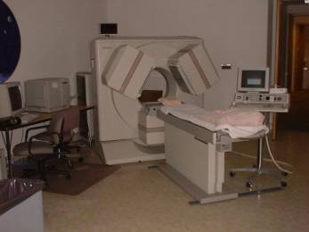
A contemporary SPECT camera (Picker Prism Triple-head model):
Although researchers are still a bit behind in proving that SPECT can confirm a diagnosis (like a chest x-ray may confirm pneumonia), my colleagues and I have found this type of brain imaging to be very useful in several ways:
Supporting the diagnosis
Along with ruling some other possible causes of psychiatric symptoms out (like brain tumors, cysts, dementia, brain trauma, vascular malformations, and stroke), SPECT imaging almost always shows brain chemical imbalances consistent with clinical symptomology.
Providing more objectivity
SPECT pictures of the brain are sensitive enough to evidence of psychological stress. For example, a person complaining of trouble concentrating, impulse control problems, or anxiety will often have corresponding activity imbalances, which I can show them easily when I go over their brain scans with them. Many people experience some relief when they discover “I’m not just making it up,” or “I can’t just snap out of it.” Furthermore, the exact measurements and their locations provide more information to use by the psychiatrist when deciding on a course of treatment. For example, SPECT imaging may save people from trying many medicines until one works, or “feeling like a guinea pig.”
Minimal risk
SPECT imaging involves injecting a small amount of radioactive compound into the vein using a small IV needle. This radioactive “tracer” compound is readily absorbed and deposits in the brain in proportion to blood flow. The radioactivity is present just long enough to take pictures, then rapidly decays. The exposure risk is typically the same as having two to three chest X-rays, well below the levels considered dangerous. SPECT imaging is approved for children as well as adults and because the tracer compound is not a dye, there is no risk of serious allergic reactions.
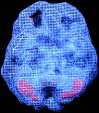
Below is a clinical example where SPECT was used in a patient and how it was helpful in both diagnosis and treatment.
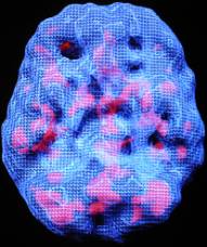
The SPECT scan on the left is of a 16-year old with depression and chronic trouble concentrating. The red areas of her brain indicate excessive activity, suggesting a more complicated mood disorder than was considered previously.
The SPECT scan on the right is of the same 16-year old after more effective treatment of her mood disorder. The medication adjustment calmed down her patchy areas of overactivity. Her depression resolved and her mood remained much more balanced.
